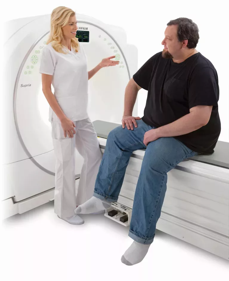
Diagnostic imaging techniques are tools that produce images of internal structures of the body to help physicians make diagnoses. The imaging methods use electromagnetic radiation and other technologies to create the images.
A wide range of diagnostic imaging tools is available to doctors. The type of tool will vary depending on the condition under evaluation.
Some of the most used diagnostic imaging tools include the following.
1. X-Ray
The x-ray is a quick and painless imaging technology that produces images of the body, especially the bones and joints.
X-rays use a low-dose radiation emitter to pass beams through your body, reflecting your internal structures on a photographic film. The body absorbs the beam of radiation differently depending on the density of a body organ. As a result, dense materials such as bones and metal will appear white on the film.
Experts perform x-rays to:
- Check the progression of a disease.
- Determine the extent of an injury.
- Evaluate how treatments are working.
X-rays are not harmful because radiation exposure levels are low. However, if you are pregnant or suspect you may be, an x-ray may not be the best option. Your doctor may recommend another imaging test, such as an ultrasound.
2. Computerized Tomography (CT) Scan
Also known as CAT scans, CT imaging technology allows doctors to see a cross-section of your body. A CT scan produces highly detailed cross-sectional images in 3D. Often, doctors will commission a CT scan when something suspicious appears on the x-ray.
The CT scan can take photos of blood vessels, body organs, soft tissue, and bones. Your doctor may recommend a CT scan to:
- Pinpoint the location of a tumor or blood clot.
- Detect internal injuries and bleeding.
- Diagnose muscle and bone disorders.
Before a CT scan, your doctor may introduce a contrast material to your body through a blood vessel, orally, or through the rectum. The contrast material helps improve the visibility of the body part under examination.
Because the images produced are of higher quality, a CT scan is relatively more expensive than x-rays or ultrasounds.
3. Magnetic Resonance Imaging (MRI)
The MRI scan is another option for cross-sectional imaging. Unlike x-ray and CT scans, MRIs use radio waves alongside strong magnetic fields to produce images. Therefore, MRIs don’t have the harmful effects of radiation.
The MRI machine is a large tube-like structure that patients travel through. The magnetic field temporarily aligns water molecules in the body. The radio waves then cause the aligned atoms to produce a signal that produces a cross-sectional image of internal organs.
MRIs are primarily used for imaging the brain and spinal cord but can also diagnose:
- Multiple sclerosis
- Disorders of the eye and inner ear
- Aneurysms of cerebral vessels
Like the CT scan, the MRI produces 3D images, albeit with better soft tissue, muscle, and fat contrast. However, MRIs take longer to administer than CT scans and x-rays.
4. Ultrasound
The ultrasound scan is also known as a sonogram. The imaging technology utilizes sound waves to produce a live video feed of internal body organs.
Before taking an ultrasound, you might be asked to drink water or fast for several hours as these may affect the quality of the images. The radiographer may administer a sedative to help you relax if necessary.
During the procedure, the Technologist applies a lubricant to the skin. An ultrasound probe translates the high-frequency sound waves to moving images displayed on a monitor.
The ultrasound scan can detect soft tissue and blood vessel concerns. Because the technique uses no radiation, it is the preferred way to view fetal development in pregnant women. Doctors also use the guidance of ultrasound in the treatment of varicose veins.
An ultrasound takes 30 minutes to an hour and generally has no known risks or side effects.
The advent of diagnostic imaging has revolutionized healthcare, allowing physicians to diagnose illnesses earlier. Moreover, the tools also reduce unnecessary invasive exploratory procedures and improve patient outcomes.
To schedule an imaging procedure with MH Imaging, call our team today.


